
Onion Cell Structure
(Middle School/High School)
What will students learn?
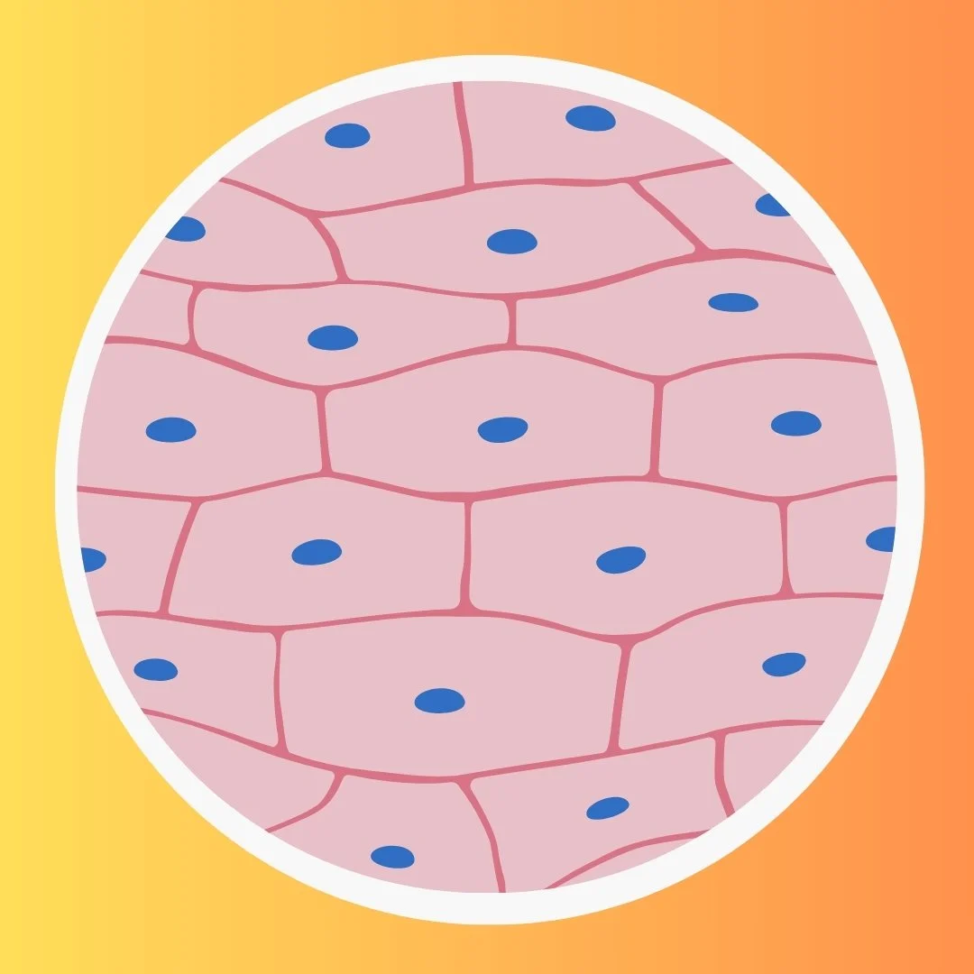
By observing onion cells under a microscope, students will learn about the basic principles of microscopy, develop skills in slide preparation, and gain insights into the structure of plant cells. This hands-on activity provides a practical understanding of cell morphology, the presence of cell walls, and the identification of cellular components such as nuclei.
What do you need?
Onion Bulb
Microscope
Microscope Slides and Coverslips
Dropper or Pipette
Staining Solution (optional)
Tweezers
Steps:
An onion is composed of layers separated by a thin membrane. For this experiment, the thin membrane will be utilized to observe onion cells.
Use tweezers to carefully peel off the thin membrane from any layer of the onion. This membrane will serve as the specimen for observation.
Add a drop of water on the microscope slide and delicately place the thin membrane on the slide.
If needed, add more drops of water on the onion peel to prevent it from drying out during observation. If you're using a staining solution, add a drop of it to the peel. Iodine is commonly used as it stains the cell components.
Gently place a coverslip over the onion peel. Ensure there are no air bubbles trapped.
Turn on the microscope and place the prepared slide on the stage.
Start with the lowest magnification objective lens (usually 4x or 10x) and bring the specimen into focus using the coarse and fine adjustment knobs.
Gradually increase the magnification using higher objective lenses (40x, 100x).
Observe the onion cells. You should be able to see rectangular cells with distinct cell walls. Look for the cell nucleus, which may appear as a darker spot within the cell.